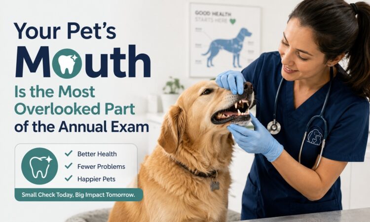
A dog with a broken leg limps. A dog with an abscessed tooth eats dinner.
That single asymmetry explains almost everything about why oral disease in pets goes untreated for years. Dogs and cats evolved to conceal weakness. Chronic mouth pain is not something they announce. They adapt, chew on one side, slow down slightly, and their owners describe them as “getting older.”
They usually aren’t. They’re usually sore.
The Prevalence Nobody Expects
Periodontal disease is the most commonly diagnosed condition in companion animals. It is not close to second place.
Veterinary literature consistently reports that 80% or more of dogs and up to 70% of cats show some stage of periodontal disease by two to three years of age. The Merck Veterinary Manual puts it at up to 80% of dogs and 70% of cats by age two. Cornell’s canine health center reports 80–90% of dogs over three.
Practice data tells the same story. One large corporate practice network found oral disease in roughly 78% of dogs and 68% of cats over the age of three, and estimated that periodontal risk climbs about 20% with each additional year of life.
And it is systematically underdiagnosed. Most pets show few outward signs until the disease is advanced.
Plaque Is the Enemy — Not Tartar
This is the most useful correction a pet owner can absorb.
That brown-gold crust on a dog’s canine tooth is calculus, or tartar. It’s ugly. It harbours bacteria. But it is not the primary driver of disease.
Plaque is. Specifically, plaque below the gumline, where you cannot see it and a toothbrush barely reaches.
The timeline is brutal:
- Plaque re-forms on clean tooth surfaces within 24 hours
- One week without home care produces gingivitis in some patients
- Three weeks without home care produces gingivitis in essentially all patients
- Periodontal pockets can become reinfected within two weeks of a professional cleaning if no home care follows
That last point reframes everything. A professional cleaning is not a solution. It is a reset. What you do in the following weeks determines whether the reset holds.
The Four Stages
| Stage | Attachment loss | What’s happening | Reversible? |
|---|---|---|---|
| 1 — Gingivitis | None | Gums inflamed, red at the margin. Bone intact. | Yes, fully |
| 2 — Early periodontitis | Under 25% | Slight bone loss visible on radiographs | Progression can be halted |
| 3 — Moderate periodontitis | 25–50% | Pockets deepen, roots exposed, mobility begins | No — management only |
| 4 — Advanced periodontitis | Over 50% | Severe bone loss, mobile or lost teeth, possible oronasal fistula | No — extraction usually required |
Here’s the part that catches owners off guard: stages are assigned per tooth, not per mouth. A dog can have healthy incisors and a stage 4 carnassial in the same visit.
Signs Worth a Phone Call
Some are obvious. Most aren’t.
Commonly noticed
- Bad breath that has become genuinely unpleasant
- Visible tartar along the gumline
- Red or bleeding gums
- Drooling, sometimes tinged pink
Frequently missed
- Chewing only on one side
- Dropping kibble, then picking it back up
- Preferring wet food after years of dry
- Pawing at the face or rubbing the muzzle on furniture
- Turning the head away when petted near the jaw
- Reduced play or fetch enthusiasm
- Sneezing or nasal discharge (a possible sign of an oronasal fistula from an upper canine)
- In cats: a chattering jaw when eating, a hallmark of tooth resorption
Absence of visible tartar means nothing. Some of the worst disease sits under clean-looking crowns.
Why Anesthesia Isn’t Optional
This deserves plain language, because the marketing around it is confusing.
“Anesthesia-free dental cleaning” — properly called non-professional dental scaling — cleans the visible crown of the tooth. It does not clean below the gumline. It does not permit probing. It does not permit radiographs.
Roughly 30% of dental disease in dogs and about 40% in cats is only visible on dental X-rays. Skipping radiographs means missing a third to nearly half of what’s wrong.
The American Veterinary Dental College’s position is that anesthesia-free dentistry is not appropriate for companion animals. The reasons are concrete:
- Sharp hand instruments in a conscious, frightened animal’s mouth risk soft tissue injury
- Without an endotracheal tube, water, debris and bacteria can be aspirated into the lungs
- The restraint required is stressful, and often the owner cannot see it happening
- The teeth look better afterward, which delays real treatment
Documented cases exist of dogs receiving anesthesia-free cleanings for years, then requiring more than a dozen extractions once someone finally took radiographs.
Clean-looking teeth are not the same as a healthy mouth.
What a Proper Dental Procedure Actually Includes
The term of art is COHAT — Comprehensive Oral Health Assessment and Treatment. Ask for it by name.
- Pre-anesthetic workup. Physical exam, bloodwork, risk assessment tailored to age and comorbidities.
- Anesthesia with intubation. Protects the airway. IV catheter, fluids, and continuous monitoring of heart rate, blood pressure, oxygenation and temperature.
- Full-mouth dental radiographs. Every tooth, every root. This is where the diagnosis actually happens.
- Charting. Probing depths, mobility, furcation exposure, gingival recession, recorded per tooth.
- Supragingival and subgingival scaling. Ultrasonic above the gumline, hand instrumentation below.
- Polishing. Smooths the microscopic scratches scaling leaves behind, which slows plaque re-adhesion.
- Treatment. Extractions, periodontal therapy, or nothing at all if the mouth is healthy.
- Recovery and a home care plan. The plan is the part that determines the next two years.
If you’re evaluating a veterinary clinic in High River, AB, ask two questions: Do you take full-mouth dental radiographs on every dental patient? and Who monitors anesthesia, and with what equipment? The answers separate a thorough practice from a cosmetic one.
Home Care, Ranked by What Actually Works
Not all prevention is equal. Here it is honestly ordered.
| Rank | Method | Effectiveness | Reality check |
|---|---|---|---|
| 1 | Daily toothbrushing | Gold standard | Every-other-day is meaningfully worse. Daily or don’t count on it. |
| 2 | VOHC-accepted dental diets | Strong | Look for the Veterinary Oral Health Council seal, not marketing language |
| 3 | VOHC-accepted chews | Good | Watch calories. Watch hardness. |
| 4 | Dental wipes / gels | Moderate | Better than nothing when brushing fails |
| 5 | Water additives | Modest | Adjunct only. Never a substitute. |
| 6 | Antlers, bones, hooves, ice | Harmful | A leading cause of slab fractures on carnassial teeth |
The rule of thumb for chews: if you can’t dent it with a thumbnail, it can crack a tooth.
Teaching a dog to accept a toothbrush
- Days 1–3: Let them lick pet toothpaste off your finger. Nothing else.
- Days 4–7: Lift the lip, touch a canine tooth with a fingertip, reward.
- Week 2: Introduce a finger brush on the outer surfaces only. Ten seconds.
- Week 3: Move to a soft brush. Thirty seconds. Outer surfaces only — the tongue handles the inside.
- Ongoing: Same time, every day. After the evening walk works well.
Never use human toothpaste. Xylitol and fluoride are not safe for pets to swallow.
The Systemic Argument
Chronic oral infection isn’t confined to the mouth. In both dogs and cats, periodontal disease has been associated with cardiac, renal, and hepatic changes, and with reduced quality of life. Some studies link it to increased mortality risk.
The mechanism is inflammation. A mouth with deep infected pockets is, in surface area terms, a persistent open wound feeding bacteria and inflammatory mediators into circulation.
Treating the mouth is not cosmetic dentistry for animals. It’s internal medicine that happens to be accessible with a probe.
Risk Factors Worth Knowing
- Small and toy breeds. Yorkshire Terriers, Toy Poodles, Dachshunds, Chihuahuas — same number of teeth, far less jaw.
- Brachycephalic breeds. Crowding and rotation trap plaque. In cats, Persians, Himalayans and Exotics show steep age-related increases in risk.
- Age. Risk rises meaningfully with every year.
- Diabetes and other immunosuppressive conditions.
- Malocclusion or retained deciduous teeth.
- Cats specifically. Tooth resorption and stomatitis are separate, painful conditions that dental radiographs catch and visual exams miss.
Frequently Asked Questions
How often should my pet have a dental cleaning? It depends on breed, size, and home care. Small breeds often need annual attention. Larger dogs with good home care may go longer. Your veterinarian charts the mouth and tells you the interval.
Is anesthesia dangerous for older pets? Age is not a disease. Modern protocols with pre-anesthetic bloodwork, IV fluids, and continuous monitoring make anesthesia safe for the large majority of senior patients. Untreated oral infection carries its own risk.
My pet still eats normally, so the mouth must be fine, right? No. Eating is a survival behaviour. Animals eat through significant pain. Appetite is one of the last things to change.
Do dental treats replace brushing? No. They help. They don’t replace. Look for the VOHC seal.
Why do cats need X-rays if their teeth look clean? Tooth resorption typically begins at or below the gumline. It’s painful and it’s common. Radiographs are the only way to catch it early.
Will my pet need extractions? Sometimes. An extracted, painful tooth is better than a retained, painful tooth. Most pets eat better within days of having diseased teeth removed.
Is bad breath in dogs normal? Mild breath, yes. Genuinely offensive breath is a clinical sign, not a personality trait.
Can I reverse periodontal disease? Stage 1 gingivitis, yes — completely. Once bone is lost, it doesn’t come back. Everything after that is containment.